
New Technique may Unravel Secrets of Biochemistry
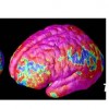
 A new paper from David Kleinfeld’s Laboratory at UC San Diego details an exciting new technique for studying biochemistry in the brain. The paper, published in the journal Nature Neuroscience, outlines a novel method for studying cell-to-cell signals that are the basis of neurotransmission. It has significant potential for uncovering the mechanisms by which many psychiatric drugs work.
What did the group find?
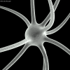
Kleinfeld’s group devised a technique that uses elaborately-named “cell-based neurotransmitter fluorescent engineered reporters” (CNiFERs for short) to examine how neurotransmitter receptors are activated. CNiFERs are cells that have been engineered to change color when acted upon by a specific neurotransmitter. The group created CNiFERs that responded to acetylcholine and implanted these cells into the frontal cortex of adult rats. When stimulated, the CNiFERs fluoresced to indicate the presence of acetylcholine in the frontal cortex.
Next, the group injected the rats with clozapine and olanzapine – neuroleptic drugs (antipsychotics) that are often used to treat schizophrenia. The implanted cells ceased to fluoresce, indicating that the drugs were blocking the transmission of acetylcholine. In other words, the group could see how neuroleptics were affecting the frontal cortex simply by looking at its color!
Why is this important?
This is very exciting and potentially very important. It is theoretically possible to create CNiFERs that respond to any neurotransmitter and to implant cells in any part of the brain. The mechanisms by which many psychiatric drugs work is largely a mystery, with selective serotonin reuptake inhibitors (SSRIs, used to treat depressions) being a case in point. With CNiFERs, researchers have a potentially powerful tool to understand how biochemical signals are relayed through the brain and the sites where they are active. This may lead to more effective treatments for disorders and lay bare many of the secrets of biochemistry that have been hidden for so long.
| Print article | This entry was posted by connolly on January 11, 2010 at 11:48 pm, and is filed under G2C Online. Follow any responses to this post through RSS 2.0. You can skip to the end and leave a response. Pinging is currently not allowed. |
No comments yet.
A new superdrug that fights pneumonia, pimples AND schizophrenia? Meet Minocycline.
about 12 years ago - No comments
A cheap drug called Minocycline, which is normally prescribed for pneumonia and acne will be tested in a new trial to reduce the symptoms of psychosis in patients suffering from schizophrenia. Schizophrenia is a mental disorder characterized by a breakdown of thought processes and by poor emotional responsiveness. According to the WHO the disorder affects…

Different sides of the same coin; twins and epigenetics
about 12 years ago - No comments
Most people are aware that monozygotic (identical) twins share the exactly the same DNA, but it might be surprising to know that traits and diseases with genetic components can vary between these twins. In the case of some psychiatric disorders with strong genetic components, there are many pairs of identical twins in which only one…
A New Neurotransmitter (D-Aspartic Acid)
about 12 years ago - No comments
In 2011, you would think that neuroscience is focused on discovering answers to high-level questions about the brain; how consciousness arises, how emotions work, what is autism, etc. Although progress is being made in all of those areas, it seems that we still have a great deal to learn about even the most basic components…

Childhood Indications of Schizophrenia
about 14 years ago - No comments
A recent 30 Year longitudinal study of individuals from New Zealand has revealed early indications of schizophrenia development later in life. Unlike many mental disorders, schizophrenia usually strikes much later in life (usually in mid to late adolescence) and so parents and patients alike may be unaware that there is a potential problem. In many…
Copy number variation in Schizophrenia
about 14 years ago - No comments
Copy number variation underlay many mental disorders

Heath Ledger’s Joker and the Hollywood Stereotype of Mental Illness
about 14 years ago - No comments
A report released this week by Dr. Peter Byrne of Newham University Hospital in London takes issue with the portrayal of mental health in Hollywood. Dr. Byrne highlights a number of characters, including Heath Ledger’s Joker from the Batman series and Jim Carrey’s character(s) in Me, Myself and Irene, which “represented a new low [for]…

Depression Genetics Suffer Major Setback
about 14 years ago - No comments
A 2003, a paper by Caspi and colleagues offered tantalizing clues about the genetics of depression, in what was widely-acclaimed as a breakthrough paper for psychiatric genetics as a whole. Now, new research by Katleeen Merikangas at the National Institute of Mental Health queries the results taking us, according to Science magazine, back to the…

Psychosis – New Study Links Gene Variant to Brain Structures
about 14 years ago - No comments
A study published in last week’s Science magazine shows how genomic science and neuroimaging can be combined to deliver insights into cognitive disorders. As well as providing an intriguing look into the neurobiology of psychosis, the study reflects a growing trend toward inter-disciplinary research in the neurosciences, What did the study show? Psychosis is a…
Schizophrenia and Autism – Opposite Ends of the Same Spectrum?
about 15 years ago - No comments
Bernard Crespi, an evolutionary geneticist at the Simon Fraser University in Burnaby, Canada, has proposed that schizophrenia and autism are the opposite ends of the same social spectrum. Speaking at the Sackler Colloquium on Evolution in Health and Medicine at the National Academy of Sciences, Crespi noted that copy number variations (CNVs) in the human…